Cholelithiasis & Biliary Colic (Gallbladder Stones / Gallstone Disease)
An authoritative clinical and educational profile of Cholelithiasis and Biliary Colic, covering cholesterol supersaturation, gallbladder hypomotility, cystic duct obstruction, constitutional homeopathic supportive management, and emergency red flags for acute ascending cholangitis (Charcot's triad), acute gallstone pancreatitis, and acute gangrenous cholecystitis.
Editorial review complete
Independent clinical validation is pending.
Quick Reference Facts
"Abdominal Ultrasound is the gold standard imaging for gallstones with >95% sensitivity; look for intraluminal echogenic foci with distinct posterior acoustic shadowing."
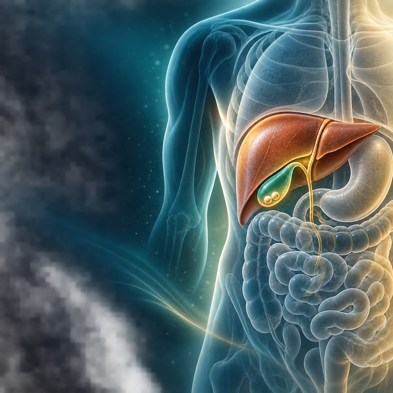
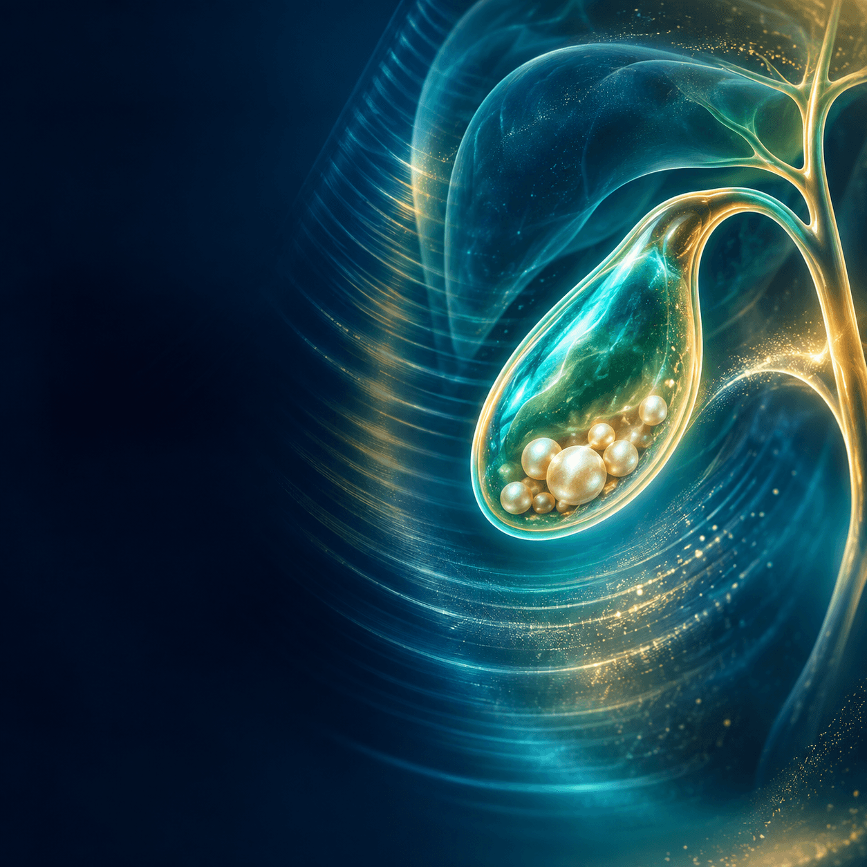
Visual guide
 Homeo Healthcare
 Homeo Healthcare
 Homeo Healthcare
In simple words
Cholelithiasis (gallbladder stones) (gallstone disease) is a very common hepatobiliary condition characterized by the formation of solid crystalline concretions (calculi) within the lumen of the gallbladder. Driven by an imbalance in biliary chemical composition (cholesterol supersaturation, gallbladder hypomotility, and accelerated crystallization), gallstones range from asymptomatic silent stones to symptomatic Biliary Colic—a severe, episodic, crescendo-decrescendo visceral pain in the epigastrium or right upper quadrant (RUQ) radiating to the right infrascapular region, typically triggered 30 to 90 minutes following a fatty, heavy meal due to transient impaction of a stone in the neck of the gallbladder (Hartmann's pouch) or cystic duct.
The presence of solid crystalline stones (cholesterol, black pigment, or brown pigment stones) inside the gallbladder, categorized clinically into asymptomatic cholelithiasis (gallbladder stones), uncomplicated symptomatic cholelithiasis (gallbladder stones) (biliary colic), and complicated gallstone disease (acute cholecystitis, choledocholithiasis, cholangitis, gallstone pancreatitis).
Common causes
- Cholesterol supersaturation: hepatic hypersecretion of cholesterol relative to solubilizing bile salts and phospholipids (lecithin), creating an unstable lithogenic bile microenvironment
- Gallbladder hypomotility and stasis: impaired gallbladder emptying and diminished cholecystokinin (CCK) receptor responsiveness allowing nucleated microcrystals to coalesce into macroscopic stones
- Accelerated cholesterol nucleation and crystallization facilitated by gallbladder mucin hypersecretion and inflammatory glycoproteins
- Pigment stone pathogenesis: Black pigment stones arise from chronic unconjugated hyperbilirubinemia (hemolytic anemias: sickle cell, thalassemia, hereditary spherocytosis, or cirrhosis); Brown pigment stones arise from chronic bacterial/parasitic biliary tract infections (E. coli, Clonorchis sinensis, Ascaris) producing bacterial beta-glucuronidase
Risk Factors
- The classic 'Female, Forty, Fertile, Fat' risk triad (female gender, age ≥40 years, multiparity/estrogen exposure, and obesity)
- Rapid, dramatic weight loss (bariatric surgery or severe very-low-calorie fasting mobilizing peripheral cholesterol and slowing gallbladder motility)
- Total Parenteral Nutrition (TPN) and prolonged fasting (absence of enteral stimulation causes profound biliary stasis)
- Medications: oral contraceptives, hormone replacement therapy (estrogen upregulates hepatic cholesterol secretion), fibrates, and ceftriaxone (biliary pseudolithiasis)
- Underlying hemolytic disorders (sickle cell disease) and terminal ileal disease (Crohn's resection impairing bile acid reabsorption)
Common symptoms
- Episodic Biliary Colic: intense, severe, steady aching or squeezing pain located in the epigastrium or right upper quadrant (RUQ), lasting from 30 minutes up to 4 to 6 hours
- Right infrascapular radiation: pain radiating around the right costal margin to the right shoulder blade, right mid-back, or interscapular region (Boeas sign)
- Postprandial exacerbation: sudden onset characteristically triggered 30 to 90 minutes following a rich, fried, or high-fat meal
- Associated symptoms: profound nausea, non-projectile vomiting, diaphoresis, restless tossing during pain paroxysms, and postprandial bloating/dyspepsia
- Asymptomatic 'silent' gallstones: >70% to 80% of individuals with gallstones remain entirely asymptomatic throughout their lifetime and are detected incidentally on routine abdominal imaging
Clinical Red Flags
Seek urgent medical attention at an emergency department or primary care clinic if you present with any of the following symptoms:
- Acute Ascending Cholangitis: Charcot's triad (high spiking fevers with chills, right upper quadrant abdominal pain, and overt jaundice) or Reynolds' pentad (adding hypotension/shock and altered mental status; life-threatening surgical emergency requiring immediate emergency biliary decompression via ERCP and IV antibiotics)
- Acute Gallstone Pancreatitis: severe, agonizing epigastric pain radiating directly through to the back, intractable vomiting, and markedly elevated serum lipase/amylase (>3 times upper limit of normal; requires emergency hospital admission and intensive resuscitation)
- Acute Cholecystitis: constant severe right upper quadrant pain lasting >6 hours, high fever, localized peritoneal guarding, and positive Murphy's sign (arrest of inspiration on deep palpation under right costal margin; requires urgent hospitalization and laparoscopic cholecystectomy)
- Gallbladder Perforation: sudden transient relief of acute cholecystitis pain followed by generalized peritonitis, abdominal rigidity, and septic shock
Lifestyle & diet support
Adopt a low-fat, high-fiber dietary pattern to avoid stimulating vigorous gallbladder contractions, eat small frequent meals at regular intervals (avoid prolonged fasting or skipping breakfast which worsens biliary stasis), avoid crash dieting and rapid weight loss (aim for a safe gradual weight loss of 0.5 to 1 kg per week), stay well hydrated, incorporate healthy unsaturated fats (extra-virgin olive oil) in moderation, and engage in daily physical exercise.
Treatment Approaches
Conventional Management
Asymptomatic gallstones generally require watchful waiting without prophylactic intervention. Symptomatic cholelithiasis (recurrent biliary colic) is definitively treated with Laparoscopic Cholecystectomy (surgical removal of the gallbladder), which is the international gold standard. Acute cholecystitis mandates urgent hospital admission, IV fluid resuscitation, broad-spectrum IV antibiotics, and early laparoscopic cholecystectomy within 24 to 72 hours of admission. Choledocholithiasis requires Endoscopic Retrograde Cholangiopancreatography (ERCP) with biliary sphincterotomy and stone extraction. Medical dissolution therapy with Oral Ursodeoxycholic Acid (UDCA) is reserved strictly for select non-surgical candidates with small (<5–10 mm), radiolucent, pure cholesterol stones and a functioning gallbladder with a patent cystic duct.
Homeopathic Approach
Homeopathic constitutional and hepatobiliary remedies (such as Chelidonium Majus, Lycopodium Clavatum, Carduus Marianus, Berberis Vulgaris, Nux Vomica, Dioscorea Villosa, Hydrastis Canadensis, Phosphorus, Chionanthus Virginica) serve as supportive care to ease postprandial biliary spasms, relieve right hypochondriac heaviness, and support digestive comfort alongside low-fat dietary moderation, ultrasound monitoring, and surgical evaluation.
Frequently Asked Questions
Clinical & academic detailShow detailHide detail
Diagnosis & tests
Investigation Protocol
Transabdominal Ultrasound (USG Abdomen) is the undisputed first-line imaging modality of choice (sensitivity and specificity >95% for detection of gallbladder stones ≥2 mm). Diagnostic sonographic criteria include: echogenic intraluminal foci casting strong posterior acoustic shadows, gravitational mobility (stones shifting positions with patient repositioning), and evaluation for complications (gallbladder wall thickening ≥3 mm, pericholecystic fluid, and sonographic Murphy's sign indicating acute cholecystitis). Magnetic Resonance Cholangiopancreatography (MRCP) or Endoscopic Ultrasound (EUS) is indicated if choledocholithiasis (common bile duct stones) is suspected.
Differential Diagnosis
Differentiate Cholelithiasis from Peptic Ulcer Disease / Perforation, Acute Pancreatitis (epigastric pain radiating straight through to the mid-back with markedly elevated lipase), Acute Appendicitis, Gastroesophageal Reflux Disease (GERD), Right-sided Nephrolithiasis (flank pain with hematuria), Sphincter of Oddi Dysfunction, and Acute Coronary Syndrome (inferior wall myocardial infarction presenting as epigastric pain).
Differential Diagnosis Matrix
Differentiate Cholelithiasis from Peptic Ulcer Disease / Perforation, Acute Pancreatitis (epigastric pain radiating straight through to the mid-back with markedly elevated lipase), Acute Appendicitis, Gastroesophageal Reflux Disease (GERD), Right-sided Nephrolithiasis (flank pain with hematuria), Sphincter of Oddi Dysfunction, and Acute Coronary Syndrome (inferior wall myocardial infarction presenting as epigastric pain).
Reference Citations & Evidence Sources
Classical Homeopathic Literature
- CIT-0007Hahnemann S.. "The Chronic Diseases: Their Peculiar Nature and Their Homoeopathic Cure." Adolph Arnold (1828).
Materia Medica & Keynotes
- CIT-0004Hahnemann S.. "Materia Medica Pura." Adolph Arnold (1811).
- CIT-0005Kent J. T.. "Lectures on Homoeopathic Materia Medica." Boericke & Tafel (1905).
- CIT-0006Boericke W.. "Pocket Manual of Homoeopathic Materia Medica." Boericke & Runyon (1901).
Clinical Reviews & Textbooks
- CIT-0023National Center for Complementary and Integrative Health. "Homeopathy: What You Need To Know." National Institutes of Health (2021).
Clinical Connections
Related Symptoms
Related Remedies
Seeking Expert Consultation?
Book an online video session with Dr. Narayan Jethwani to get an individualized constitutional homeopathic protocol tailored to your case.
Medical Safety Disclaimer
All content on the Homeo Healthcare platform is strictly for educational purposes and is not personal medical advice, diagnosis, or treatment. Homeopathic remedy considerations are provided for clinician review or require individualized consultation with a qualified physician. Never delay seeking professional medical advice or emergency medical care due to content you have read on this website.